
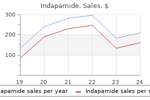
Only $0.56 per item
Indapamide dosages: 2.5 mg, 1.5 mg
Indapamide packs: 30 pills, 60 pills, 90 pills, 120 pills, 180 pills, 270 pills, 360 pills
In stock: 831
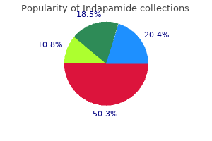
10 of 10
Votes: 20 votes
Total customer reviews: 20
Description
Stem cell transplantation using tissue harvested from the fellow eye of the patient may eventually be required blood pressure medication good for pregnancy purchase 2.5 mg indapamide. Once removed from the ocular surface, the lesion can be placed on filter paper and the edges labeled to facilitate histopathologic diagnosis. Care should be taken to avoid damage to the specimen during removal, as any damage could make the lesion more difficult to interpret. Diagnosis of conjunctival tumors can be challenging, especially for general pathologists. Topical Chemotherapy Topical chemotherapy can be used in the management of ocular surface tumors as primary treatment before or instead of surgical tumor removal or, more commonly, as an adjunctive therapy after tumor excision. Recent reports have suggested the use of topical interferon-2b as primary therapy for tumors in the squamous cell family; however, one potential disadvantage of this course of treatment is the lack of histopathologic diagnosis. In addition, the effectiveness of topical interferon-2b in the treatment of melanotic tumors has not been proven. Since there is some overlap in the appearance of various tumors (eg, amelanotic conjunctival melanoma may resemble squamous cell carcinoma), there is concern that if chemotherapy is elected and the tumor does not respond, valuable time in the management of a potentially deadly lesion may have been lost. Another disadvantage of topical interferon-2b is that it is expensive and may take several months to work. Some authors advocate using topical interferon-2b before definitive surgical removal, with the purpose of trying to shrink large ocular surface squamous tumors preoperatively. The optimal topical chemotherapy regimen has not been determined in controlled studies, but, typically, topical interferon-2b is given 4 times daily until clinical response occurs, usually within 2 to 4 months. Placement of punctal plugs reduces the chance of systemic absorption and helps prevent punctal stenosis. Once a tumor has been managed, long-term, regular follow-up is essential, because malignant conjunctival tumors can recur. Complete examination of the ocular surface and palpation of regional lymph nodes should be performed at each visit. Patients with malignant ocular surface tumors should be referred to a dermatologist for a complete skin evaluation. The remainder of this chapter will focus on the clinical characteristics of various benign and malignant tumors of the ocular surface. Topical interferon or surgical excision for the management of primary ocular surface squamous neoplasia. Tumors of Epithelial Origin Table 8-1 lists the epithelial tumors of the conjunctiva and cornea.
Vitamin O. Indapamide.
- How does Vitamin O work?
- Arthritis; asthma; constipation; depression; diabetes; dizziness; headaches; increasing energy; improving alertness, concentration, immune function, and memory; irritability; lung disease; menopause; mouth sores; muscle aches and pains; obesity; premenstrual syndrome; sexual problems; and many other uses.
- Dosing considerations for Vitamin O.
- Are there safety concerns?
- What is Vitamin O?
Source: http://www.rxlist.com/script/main/art.asp?articlekey=96461
B blood pressure medication what does it do purchase indapamide 2.5 mg amex, C, increasing recognition that Mycobacterium Fluorescein angiogram images showing early tuberculosis can cause inflammation that also simulates hypofluorescence of the active area followed by typical serpiginous choroiditis. D, Fundus autofluorescence image demonstrating marked associated disease has been called multifocal autofluorescence in the areas of disease serpiginoid choroiditis or serpiginous-like activity (arrows). The lesions tend to appear and progress in multiple areas rather than to spread out centrifugally as they do in serpiginous choroiditis. Patients with serpiginous-like choroiditis exhibit a more prominent inflammatory cellular reaction in the vitreous than do patients with serpiginous choroiditis. Presumed tubercular serpiginouslike choroiditis: clinical presentations and management. For this discussion, the traditional nomenclature is used to discuss the 3 related diseases as discrete entities. Symptoms include floaters, photopsias, enlargement of the physiologic blind spot, and decreased vision. Acute lesions have a creamier, opaque appearance that becomes punched out over time. Fluorescein angiography shows early hypofluorescence with late staining of acute active lesions, whereas atrophic lesions behave as transmission defects (early hyperfluorescence that fades in the late phases of the angiogram). A viral etiology involving herpes simplex and Epstein-Barr virus has been postulated, but neither has been conclusively demonstrated. B, Late staining the diagnosis is one of exclusion, as many other of lesions (arrows). The visual prognosis is guarded, with permanent vision loss in at least 1 eye occurring in up to 75% of patients as a result of the complications associated with chronic, recurrent inflammation. In one study, the incidence of vision loss to 20/200 or worse was 12% per eye-year in the affected eye. The intravitreal fluocinolone acetonide implant is also a potential treatment option. However, active inflammation can stimulate neovascularization and blunt the effects of these treatments, so it is important that any inflammatory component be well controlled. Redefining multifocal choroiditis and panuveitis and punctate inner choroidopathy through multimodal imaging. Multifocal choroiditis with panuveitis: incidence of ocular complications and loss of visual acuity. C, Three years later, fundus photograph shows multiple visible spots (arrowheads) corresponding to previously visualized hypoautofluorescent spots. Multifocal choroiditis with panuveitis and punctate inner choroidopathy: comparison of clinical characteristics at presentation. Clinical features and incidence rate of ocular complications in punctate inner choroidopathy.

Specifications/Details
The most commonly used gonioscopic grading systems are the Shaffer and Spaeth systems arrhythmia consultants buy indapamide 1.5 mg low price. If a grading system is used, the clinician should specify which system is being used. The Shaffer system describes the angle between the trabecular meshwork and the iris as follows: Grade 4: the angle between the iris and the surface of the trabecular meshwork is 45°. Grade 3: the angle between the iris and the surface of the trabecular meshwork is greater than 20° but less than 45°. Grade 2: the angle between the iris and the surface of the trabecular meshwork is 20°. Grade 1: the angle between the iris and the surface of the trabecular meshwork is 10°. Slit: the angle between the iris and the surface of the trabecular meshwork is less than 10°. Pathologic causes include hypotony and elevated episcleral venous pressure, as in carotid-cavernous fistula or SturgeWeber syndrome. Normal blood vessels in the angle include radial iris vessels, portions of the arterial circle of the ciliary body, and vertical branches of the anterior ciliary arteries. Normal vessels are oriented either radially along the iris or circumferentially (in a serpentine manner) in the ciliary body face. The vessels seen in Fuchs classification of the anterior chamber angle, heterochromic uveitis are fine, branching, unsheathed, based on 3 variables: angular width of the angle and meandering. Patients with neovascular glaucoma recess (A); configuration of the peripheral iris (B); and apparent insertion of the iris root (C). The pigmentation pattern of an individual angle is dynamic over time, especially in conditions such as pigment dispersion syndrome. Note the red line posterior to the trabecular meshwork in this patient with or pseudoexfoliation syndrome. Pseudoexfoliation elevated episcleral venous pressure resulting in syndrome may appear clinically similar to pigment blood reflux into the Schlemm canal. In addition, a line of pigment deposition anterior to the Schwalbe line is often present in pseudoexfoliation syndrome (Sampaolesi line). Other conditions that cause increased anterior chamber angle pigmentation include melanoma, trauma, surgery, inflammation, angle closure, and hyphema. Posttraumatic angle recession may be associated with monocular open-angle glaucoma. If the ciliary body separates from the scleral spur (cyclodialysis), it will appear gonioscopically as a deep angle recess with a gap between the scleral spur and the ciliary body. For further discussion of retinal involvement in the visual process, see Section 12, Retina and Vitreous.
Syndromes
- What other symptoms do you have -- headaches, abdominal pain, breast tenderness, dry mouth, excessive thirst, unintended weight loss?
- Provide picture books
- Congenital cytomegalovirus
- Simple -- not affecting awareness or memory
- Pyrazinamide
- Heavy, prolonged, or irregular menstrual periods
- Shortness of breath
- Heart rhythm problems, such as atrial fibrillation or paroxysmal supraventricular tachycardia (PSVT) that began recently or that cannot be controlled with medicines may be treated this way.
The chemical makeup of the secretion blood pressure levels buy 1.5 mg indapamide mastercard, as well as the regulation of the secretion, varies remarkably from organ to organ. In the oral cavity, secretory and absorptive activities of the acinar and ductule cells of the salivary glands result in the production of a large volume, compared with glandular weight, of hypotonic solution that is rich in potassium and bicarbonate compared with plasma. Through a secondary active transport system involving apical membrane chloride channels, acinar cells secrete a solution similar in inorganic composition to that of plasma. As this fluid passes through the ducts, sodium and chloride are absorbed in partial exchange for potassium and bicarbonate to result in the final secretion. In addition to the inorganic components, several of the salivary glands contain cells that secrete amylase and lipase, which begin the process of digestion; lysozyme and lactoferrin, which have antimicrobial activity; and mucus. These components are synthesized in the endoplasmic reticulum and stored in secretory granules until they are released. Regulation of salivary secretion is done almost entirely through neurocrine pathways, primarily parasympathetic. However, salivary glands are among the few organs whose secretions are stimulated by both parasympathetic and sympathetic divisions of the autonomic nervous system. In the body of the stomach, parietal (oxyntic), chief (peptic), and mucus neck cells secrete hydrochloric acid, pepsin, and soluble mucus, respectively. In addition, surface epithelial cells in all regions secrete a more viscous mucus in an alkaline fluid. All of these products are secreted into the gastric lumen to take part in digestion of ingested foodstuffs, and in protection of the stomach. In addition to these digestive and protective secretions, the parietal cells secrete intrinsic factor which is necessary for the absorption of vitamin B12. In the antrum, there are endocrine (G) cells that secrete gastrin in an endocrine fashion to stimulate parietal cells, and endocrine cells that secrete somatostatin in a paracrine fashion to inhibit G cell secretion of gastrin. Upon stimulation of the parietal cell, the tubulovesicles fuse with the apical cell membrane, which contains potassium channels. During the secretion of this acid into the gastric lumen, bicarbonate is produced and released into the venous blood, producing an alkaline tide. Secretion of the pepsin precursor pepsinogen by chief cells is via exocytosis of protein that has been synthesized in the endoplasmic reticulum and stored in secretory granules. In the presence of acid in the lumen of the stomach, the secreted pepsinogen is converted to the active protease pepsin. These cells, which are adjacent to parietal cells, release histamine, which in turn acts in a paracrine manner to stimulate acid secretion. Upon stimulation, gastrin is released into the bloodstream and circulates back to the stomach to act on parietal cells.
Related Products
Additional information:
Usage: b.i.d.
Tags: cheap indapamide 2.5 mg with visa, order indapamide 1.5 mg with mastercard, buy 1.5 mg indapamide free shipping, indapamide 2.5 mg purchase without prescription
Customer Reviews
Real Experiences: Customer Reviews on Lozol
Tom, 44 years: As the wound heals, erosions may occur along the sutures, leading to eye rubbing, epithelial defects, vascularization, and mucus accumulation.
Sobota, 29 years: The racial or ethnic background of the patient is relevant, as conjunctival pigment may be normal in some patients (eg, those with pigmented skin) but more worrisome in others.
Achmed, 40 years: Wessely rings may persist for some time in corneas traumatized by a foreign body, even after the foreign body is removed.
Pranck, 49 years: Endoscopic cyclodestruction is used in eyes with better visual potential because there is less damage to the ciliary body.
Bernado, 38 years: Estrogen and progesterone replacement and adequate calcium and vitamin D intake are indicated.
Marlo, 32 years: In the small intestine, potassium is concentrated in the chyme as sodium and water are absorbed, but because the paracellular pathways are "leaky," potassium is absorbed passively down its electrochemical gradient.