
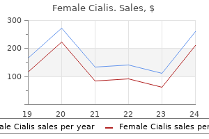
Only $0.8 per item
Female Cialis dosages: 20 mg, 10 mg
Female Cialis packs: 30 pills, 60 pills, 90 pills, 120 pills, 180 pills, 270 pills, 360 pills
In stock: 962
9 of 10
Votes: 338 votes
Total customer reviews: 338
Description
The anterior sagittal cut through the liver is made in the plane of the fossa for the gallbladder menopause urinary frequency buy female cialis 20 mg mastercard, and the posterior sagittal cut is in the plane of the fissure for the ligamentum venosum. These cuts have been joined by a narrow coronal cut in the plane of the porta hepatis. The relationship of the liver to the anterior (intraperitoneal) abdominal viscera is demonstrated. The portal triad passes between the layers of the hepatoduodenal ligament to enter the 1162 liver at the porta hepatis. The common hepatic artery passes between the layers of the hepatogastric ligament. The essentially midline plane defined by the attachment of the falciform ligament and the left sagittal fissure separates a large right lobe from a much smaller left lobe. On the slanted visceral surface, the right and left sagittal fissures course on each side of-and the transverse porta hepatis separates-two accessory lobes (parts of the anatomic right lobe): the quadrate lobe anteriorly and inferiorly and the caudate lobe posteriorly and superiorly. The caudate lobe was so-named not because it is caudal in position (it is not) but because it often gives rise to a "tail" in the form of an elongated papillary process. Each part receives its own primary branch of the hepatic artery and hepatic portal vein and is drained by its own hepatic duct. The liver can be further subdivided into four divisions and then into eight surgically resectable hepatic segments, each served independently by a secondary or tertiary branch of the portal triad, respectively. The right, intermediate, and left hepatic veins course within three planes or fissures [right portal (R), main portal (M), and umbilical (U)] that divide the liver into four vertical divisions, each served by a secondary (2°) branch of the portal triad. Three divisions are 1165 further subdivided at the transverse portal plane (T) into hepatic segments, each supplied by tertiary (3°) branches of the triad. The hepatic veins are intersegmental, draining the portions of multiple segments adjacent to them. Injection of latex into the right (red) and left (blue) portal veins demonstrates the right and left livers and the Cantlie line that demarcates them on the diaphragmatic surface. Each part, division, and segment has an identifying name; segments are also identified by Roman numerals. Under the schema of the previous terminology, the caudate lobe was divided into right and left halves. Except for the caudate lobe (segment I), the liver is divided into right and left livers based on the primary (1°) division of the portal triad into right and left branches, the plane between the right and the left livers being the main portal fissure in which the middle hepatic vein lies. The right and left livers are subdivided vertically into medial and lateral divisions by the right portal and umbilical fissures, in which the right and left hepatic veins lie. The caudate lobe (segment I, bringing the total number of segments to eight) is supplied by branches of both divisions and is drained by its own minor hepatic veins. While the pattern of segmentation described here is the most common pattern, the segments vary considerably in size and shape as a result of individual variation in the branching of the hepatic and portal vessels.
Hexacosanol (Policosanol). Female Cialis.
- Are there any interactions with medications?
- Dosing considerations for Policosanol.
- Are there safety concerns?
- High cholesterol, inherited high cholesterol (familial hypercholesterolemia), intermittent claudication, increasing blood flow to the heart in people with coronary heart disease, and other conditions.
- What is Policosanol?
- How does Policosanol work?
Source: http://www.rxlist.com/script/main/art.asp?articlekey=96177
Inferiorly menopause yellow discharge female cialis 20 mg lowest price, the fascia splits into two layers, which attach to the lateral and medial surfaces of the zygomatic arch. When it contracts, exerting a strong downward pull on the zygomatic arch, the temporal fascia provides resistance. In this superficial dissection of the great muscles on the side of the cranium, the parotid gland and most of the temporal fascia have been removed. The facial artery passes deep to the submandibular gland, whereas the facial vein passes superficial to it. Infratemporal Fossa the infratemporal fossa is an irregularly shaped space deep and inferior to the zygomatic arch, deep to the ramus of the mandible, and posterior to the maxilla. It communicates with the temporal fossa through the interval between (deep to) the zygomatic arch and (superficial to) the cranial bones. Superiorly: the inferior (infratemporal) surface of the greater wing of the sphenoid. Inferiorly: where the medial pterygoid muscle attaches to the mandible near its angle. The sphenomandibular ligament passively bears the weight of the lower jaw and is the "swinging hinge" of the mandible, permitting protrusion and retrusion as well as elevation and depression. The parotid and temporal regions and the infratemporal fossa collectively include the temporomandibular joint and the muscles of mastication that produce its movements. The bony articular surfaces involved are the mandibular fossa and articular tubercle of the temporal bone superiorly and the head of the mandible inferiorly. The loose fibrous layer of the joint capsule attaches to the margins of the articular cartilage on the temporal bone and around the neck of the mandible. This creates separate superior and inferior articular cavities, or compartments, lined by separate superior and inferior synovial membranes. The gliding movements of protrusion and retrusion (translation) occur between the temporal bone and the articular disc (superior cavity). The hinge movements of depression and elevation and the rotational or pivoting movements occur in the inferior compartment. Two extrinsic ligaments and the lateral ligament connect the mandible to the cranium. The stylomandibular ligament, which is actually a thickening of the fibrous capsule of the parotid gland, runs from the styloid process to the angle of the mandible. The sphenomandibular ligament runs from the spine of the sphenoid to the lingula of the mandible. When the mouth is closed and at rest, the heads of the mandible are held in the retracted position in the mandibular fossae, and the chin is elevated by the tonus of the retractors and elevators of the mandible. When sleeping in the supine or sitting position (head upright), as one 2086 enters a state of deep sleep, the tonic contraction relaxes and gravity causes depression of the mandible (the mouth falls open). To enable more than a small amount of depression of the mandible-that is, to open the mouth wider than just to separate the upper and lower teeth-the head of the mandible and articular disc must move anteriorly on the articular surface until the head lies inferior to the articular tubercle (a movement referred to as "translation" by dentists).
Specifications/Details
Injury to Nerves Supplying Eyelids Because it supplies the levator palpebrae superioris breast cancer chemotherapy drugs 20 mg female cialis order fast delivery, a lesion of the oculomotor nerve causes paralysis of the muscle, and the superior eyelid droops (ptosis). The loss of tonus of the muscle in the inferior eyelid causes the lid to fall away (evert) from the surface of the eyeball, leading to drying of the cornea. Thus, irritation of the unprotected eyeball results in excessive but inefficient lacrimation (tear formation). Excessive lacrimal fluid also forms when the lacrimal drainage apparatus is obstructed, thereby preventing the fluid from reaching the inferior part of the eyeball. People often dab their eyes constantly to wipe the tears, resulting in further irritation. Inflammation of Palpebral Glands Any of the glands in the eyelid may become inflamed and swollen from infection or obstruction of their ducts. If the ducts of the ciliary glands are obstructed, a painful red suppurative (pus-producing) swelling, a sty (hordeolum), develops on the eyelid. Obstruction of a tarsal gland produces inflammation, a tarsal chalazion, that protrudes toward the eyeball and rubs against it as the eyelids blink. Hyperemia of Conjunctiva the conjunctiva is colorless, except when its vessels are dilated and congested ("bloodshot eyes"). An inflamed conjunctiva, conjunctivitis ("pinkeye"), is a common contagious infection of the eye. Subconjunctival Hemorrhages 2059 Subconjunctival hemorrhages are common and are manifested by bright or dark red patches deep to and within the bulbar conjunctiva. A blow to the eye, excessively hard blowing of the nose, and paroxysms of coughing or violent sneezing can cause hemorrhages resulting from rupture of small subconjunctival capillaries. Development of Retina the retina and optic nerve develop from the optic cup, a cup-like outgrowth of the embryonic forebrain, the optic vesicle. Hence, the optic nerve is invested with cranial meninges and an extension of the subarachnoid space. The central retinal artery and vein cross the subarachnoid space and run within the distal part of the optic nerve. The pigment cell layer of the retina develops from the outer layer of the optic cup, and the neural layer develops from the inner layer of the cup (Moore et al. Although the pigment cell layer becomes firmly fixed to the choroid, its attachment to the neural layer is not firm. A detached retina usually results from seepage of fluid between the neural and pigment cell layers of the retina, perhaps days or even weeks after trauma to the eye. Persons with a retinal detachment may complain of flashes of light or specks floating in front of the eye. Pupillary Light Reflex the pupillary light reflex is tested using a penlight during a neurological examination. When light enters one eye, both pupils constrict because each retina sends fibers into the optic tracts of both sides.
Syndromes
- Stress
- Diagnose a fracture, when it cannot be seen on a regular x-ray (most commonly hip fractures, stress fractures in the feet or legs, or spine fractures)
- Sudden, severe pain anywhere in the body
- Fumes from burning coal converted into organic mercury by certain organisms
- Osteomyelitis (inflammation of the bone caused by an infection)
- Vitamin B1 (thiamine)
The internal pudendal artery leaves the gluteal region immediately by crossing the ischial spine/sacrospinous ligament and enters the perineum through the lesser sciatic foramen menstruation on the pill 10 mg female cialis purchase. There are usually four perforating arteries of the 1680 profunda femoris artery, three arising in the anterior compartment and the fourth being the terminal branch of the profunda femoris artery itself. The perforating arteries are large vessels, unusual in the limbs for their transverse, intercompartmental course. Surgeons operating in the posterior compartment are careful to identify them to avoid inadvertent injury. They perforate the aponeurotic portion of the distal attachment of the adductor magnus to enter the posterior compartment. Within the posterior compartment, they typically give rise to muscular branches to the hamstrings and anastomotic branches that ascend or descend to unite with those arising superiorly or inferiorly from the other perforating arteries or the inferior gluteal and popliteal artery. A continuous anastomotic arterial chain thus extends from the gluteal to popliteal regions, which gives rise to additional branches to muscles and to the sciatic nerve. After giving off their posterior compartment branches, the perforating arteries pierce the lateral intermuscular septum to enter the anterior compartment, where they supply the vastus lateralis muscle. The superior and inferior gluteal veins accompany the corresponding arteries through the greater sciatic foramen, superior and inferior to the piriformis, respectively. They communicate with tributaries of the femoral vein, thereby providing alternative routes for the return of blood from the lower limb. Lymph from the deep tissues of the gluteal region enters the pelvis along the gluteal veins, draining to the superior and inferior gluteal lymph nodes; from them, it passes to the iliac and lateral lumbar (caval/aortic) lymph nodes. Lymph from superficial tissues of the gluteal region passes initially to the superficial inguinal nodes, which also receive lymph from the thigh. Lymph from all the superficial inguinal nodes passes via efferent lymph vessels to the external and common iliac, and right and left lumbar (caval/aortic) lymph nodes, draining via lumbar lymphatic trunks to the cisterna chyli and thoracic duct. The internal pudendal veins accompany the internal pudendal arteries and join to form a single vein that enters the internal iliac vein. Perforating veins accompany the arteries of the same name to drain blood from the posterior compartment of the thigh into the profunda femoris vein. The perforating veins, like the arteries, usually communicate inferiorly with the popliteal vein and superiorly with the inferior gluteal vein. Lymph from the superficial tissues of the gluteal region enters the superficial inguinal lymph nodes, which also receive lymph from the thigh. All the superficial inguinal nodes send efferent lymphatic vessels to the external iliac lymph nodes. In terms of the vascular supply to the lower limb as a whole, the majority of the arterial blood coming to the limb and most of the venous blood and lymph exiting from it pass along the more protected anteromedial aspect of the limb. Surface Anatomy of Gluteal and Posterior Thigh Regions the skin of the gluteal region is usually thick and coarse, especially in men, whereas the skin of the thigh is relatively thin and loosely attached to the underlying subcutaneous tissue. A line joining these dimples, often more visible in women than in men, passes through the S2 spinous process, indicating the level of the lowest limit of the dural sac.
Related Products
Additional information:
Usage: p.c.
Tags: 10 mg female cialis mastercard, quality 20 mg female cialis, generic 20 mg female cialis free shipping, buy female cialis 10 mg visa
Customer Reviews
Real Experiences: Customer Reviews on Female Cialis
Bengerd, 52 years: Renal Transplantation Renal transplantation is the preferred treatment for selected cases of chronic renal failure. The atria are receiving chambers that pump blood into the ventricles (the discharging chambers). These muscles appear to have a weak expiratory function and may also provide proprioceptive information. If labor is extended or the level of anesthesia is inadequate, it may be difficult or impossible to re-administer the anesthesia.
Curtis, 35 years: The arch of the aorta (20) is obliquely placed (more sagittal than transverse), with the ascending end anteriorly in the midline, and the descending end posteriorly and to the left of the vertebral bodies (17). The repeated depolarizations also exhaust the marginal energy supplies within the penumbra, which may transform from non-lethal to irreversible injury. The posterior intercostal arteries: of the 1st and 2nd intercostal spaces arise from the supreme (superior) intercostal artery, a branch of the costocervical trunk of the subclavian artery. Blood received by the confluence of sinuses is drained by the transverse sinuses, but rarely equally.
Larson, 47 years: The recurrent laryngeal nerves arise from the vagus nerves in the inferior part of the neck. The subarachnoid space around 2017 the optic nerve is continuous with the space between the arachnoid and the pia covering the brain. There are also many minute openings of the ducts of mucussecreting urethral glands into the spongy urethra. All internal surfaces not covered with or made of articular cartilage (blue or gray in the case of the menisci) are lined with synovial membrane (mostly purple, but transparent and colorless where it is covering nonarticular surfaces of the femur).
Daro, 29 years: This is one of the most common injuries to the hip region, usually occurring in association with collision sports, such as the various forms of football, ice hockey, and volleyball. White matter oedema or discrete white matter lesions are present in about two-thirds of cases. The two layers of peritoneum forming the falciform ligament separate over the superior aspect of the liver to form the anterior layer of the coronary ligament, leaving the bare area of the liver without a peritoneal covering. On the slanted visceral surface, the right and left sagittal fissures course on each side of-and the transverse porta hepatis separates-two accessory lobes (parts of the anatomic right lobe): the quadrate lobe anteriorly and inferiorly and the caudate lobe posteriorly and superiorly.