
Only $0.61 per item
Azulfidine dosages: 500 mg
Azulfidine packs: 30 pills, 60 pills, 90 pills, 120 pills, 180 pills, 270 pills
In stock: 797
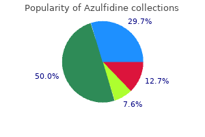
9 of 10
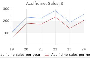
Votes: 143 votes
Total customer reviews: 143
Description
Lemniscal fibres relay only in the central nucleus pain treatment pancreatitis buy cheap azulfidine 500 mg, and some pass without relay to the medial geniculate nucleus. In humans, the ventral division of the medial geniculate nucleus receives a topographic projection from the central nucleus and the dorsal division receives a similar projection from the dorsal cortex. Some colliculogeniculate fibres do not relay in the geniculate nucleus but continue, with those that do, via the auditory radiation to the auditory cortex. A descending projection from the auditory cortex reaches the inferior colliculus via the medial geniculate nucleus. This descending path may produce effects at levels from the medial geniculate nucleus downwards, and it probably links with efferent cochlear fibres, through the superior olivary and cochlear nuclei. It is in line with the ventromedial part of the oculomotor nucleus, in the position of the somatic efferent column. The trochlear nucleus is caudal to the oculomotor nucleus and distinguished by the smaller size of its neurones. The afferent inputs to the trochlear nucleus are similar to those described for the oculomotor nucleus. Trochlear efferent fibres pass laterodorsally round the central grey matter, descending caudally medial to the mesencephalic nucleus as they do so, to reach the upper end of the superior medullary velum, where they decussate and emerge lateral to the frenulum and caudal to the inferior colliculus. It ascends to the interstitial nucleus of Cajal, which lies in the lateral wall of the third ventricle, just above the cerebral aqueduct. The fasciculus retains its position relative to the central grey matter through the midbrain, pons and upper medulla, but is displaced ventrally by the motor (pyramidal) decussation containing corticospinal fibres. The medial longitudinal fasciculus interconnects the oculomotor, trochlear, abducens, EdingerWestphal preganglionic, vestibular, reticular and spinal accessory nuclei, coordinating conjugate eye movements and associated movements of the head and neck. Those from the superior nucleus remain uncrossed, while the others are partly crossed. Some fibres reach the interstitial and posterior commissural nuclei, and some decussate to the contralateral nuclei. Descending axons, from the medial vestibular nuclei and perhaps the lateral and inferior nuclei, partially decussate and descend in the fasciculus to form the medial vestibulospinal tract, which travels in the medial longitudinal fasciculus into the ventral funiculus of the spinal cord (see Ch. Fibres join the fasciculus from the dorsal trapezoid, lateral lemniscal and posterior commissural nuclei, which means that both the cochlear and vestibular components of the vestibulocochlear nerve may influence movements of the eyes and head via the medial longitudinal fasciculus. Some vestibular fibres may ascend in the medial longitudinal fasciculus as far as the thalamus. It has a central, ovoid, main nucleus, which is lateral to the periaqueductal grey matter. It is surrounded by a lamina of nerve fibres, many from the lateral lemniscus, which terminate in it.
Common Ash (Ash). Azulfidine.
- Are there safety concerns?
- Dosing considerations for Ash.
- What is Ash?
- How does Ash work?
- Fever, arthritis, gout, bladder complaints, constipation, increasing urine production (diuretic), and other conditions.
Source: http://www.rxlist.com/script/main/art.asp?articlekey=96308
The lateral stria treating pain in dogs hips azulfidine 500 mg buy overnight delivery, clothed by the lateral olfactory gyrus, and the intermediate stria (when present) terminate in the rostral parts of the piriform area, including the olfactory trigone and tubercle, anterior perforated substance, uncus and entorhinal area of the anterior part of the future parahippocampal gyrus. The forward growth of the temporal pole and the general expansion of the neocortex cause the lateral olfactory gyrus to bend laterally, the summit of the convexity lying at the anteroinferior corner of the developing insula. During the fourth and fifth months, much of the piriform area becomes submerged by the adjoining neocortex and, in the adult, only a part of it remains visible on the inferior aspect of the cerebrum. The limbic lobe is the first part of the cortex to differentiate and, at first, it forms a continuous, almost circular, strip on the medial and inferior aspects of the hemisphere. Below and in front, where the stalk of the olfactory tract is attached, it constitutes a part of the piriform area. In this region, the neural progenitors of the developing cortex proliferate and migrate. The wall of the hemisphere thickens, producing an elevation that projects into the medial side of the ventricle. The parieto-occipital sulcus appears at about that time on the medial aspect of the hemisphere. Its appearance seems associated with an increase in the number of splenial fibres in the corpus callosum. Over the same period, the posterior part of the calcarine sulcus appears as a shallow groove extending forwards from a region near the occipital pole. It is a true infolding of the cortex in the long axis of the striate area and produces an elevation, the calcar avis, on the medial wall of the posterior horn of the ventricle. During the fifth month, the cingulate sulcus appears on the medial aspect of the hemisphere, and sulci appear on the inferior and superolateral aspects in the sixth month. The central, precentral and postcentral sulci appear, each in two parts, upper and lower, which usually coalesce shortly afterwards, although they may remain discontinuous. The superior and inferior frontal, the intraparietal, occipital, superior and inferior temporal, occipitotemporal, collateral and rhinal sulci all make their appearance during the same period. At the time of their appearance, the two hemispheres are connected to each other by the median part of the telencephalon. The roof plate of this area remains epithelial, while its floor becomes invaded by the decussating fibres of the optic nerves and developing hypothalamic nuclei. These two routes are thus not available for the passage of commissural fibres passing from hemisphere to hemisphere across the median plane, and these fibres therefore pass through the rostral wall of the interventricular foramen, i. The first commissures to develop are those associated with the palaeocortex and archicortex. In addition, the two hippocampi become interconnected by transverse fibres that cross from fornix to fornix in the upper part of the lamina terminalis as the commissure of the fornix (hippocampal commissure). Various other decussating fibre bundles (known as the supraoptic commissures, although they are not true commissures) develop in the lamina terminalis immediately dorsal to the optic chiasma, between it and the anterior commissure.
Specifications/Details
However pain heel treatment buy azulfidine 500 mg overnight delivery, venoconstriction is an important component of the baroreceptor reflex because it reduces vein compliance and therefore capacity, effectively mobilizing blood to maintain or increase central venous pressure, and hence cardiac output. Vasoconstriction in cutaneous veins in response to cooling is important in thermoregulation. A valve is formed by an inward projection of the intima, strengthened by collagen and elastic fibres, and covered by endothelium that differs in orientation on its two surfaces. Surfaces facing the vessel wall have transversely arranged endothelial cells, whereas on the luminal surface of the valve, over which the main stream of blood flows, cells are arranged longitudinally in the direction of flow. Most commonly two, or occasionally three, valves lie opposite one another; sometimes only one is present. They are found in small veins or where tributaries join Venules When two or more capillaries converge, the resulting vessel is larger (1030 µm) and is known as a venule (postcapillary venule). Elsewhere, venules are believed to be a major site where migration of neutrophils, macrophages and other leukocytes into extravascular spaces occurs, and where neutrophils may temporarily attach, forming marginated pools. In general, the endothelium of venules has few tight junctions and is relatively permeable. The intercellular junctions of venules are sensitive to inflammatory agents, which increase their permeability to fluids and defensive cells, and facilitate leukocyte extravasation by diapedesis. Venules do not acquire musculature until they are about 50 µm in outer diameter, when they are known as muscular venules. This distinction is important because postcapillary venules, which lack muscle in their walls, are as permeable to solutes as capillaries and are thus part of the microcirculatory bed. At the level of the postcapillary venule the cross-sectional area of the vascular tree is at its maximum, and there is a dramatic fall in pressure (from 25 mmHg in the capillary to approximately 5 mmHg). Muscular venules converge to produce a series of veins of progressively larger diameter. Femoral vein Fluid exchange in the microvasculature Orifice of tributary the microvasculature is important for the creation and maintenance of the interstitial fluid that bathes the cells. The thin walls of capillaries and small venules allow easy diffusion of fluid and most small molecules, but the endothelial barrier prevents movement of proteins; consequently, plasma and interstitial fluid have almost identical compositions, except that the latter contains very little protein. Fluid transfer across these exchange vessels is driven by the balance between the hydrostatic pressure. Inflammation and endothelial permeability Inflammatory mediators increase the permeability of capillaries and small venules by causing contraction of endothelial cells and so loosening tight junctions. This facilitates leukocyte extravasation by diapedesis but also disrupts normal barrier function, allowing extravasation of protein and fluids. The consequence is tissue oedema and the swelling that is commonly associated with inflammation.
Syndromes
- Vomiting
- Certain medical conditions such as diabetes
- Red, flushed face
- If the medication was prescribed for the patient
- Blisters or ulcers -- most often on the mouth, lips and gums, or genitals
- Catheter that is blocked or that has a kink in it
- Call the local emergency number, such as 911.
- Sleep disorders
- Nausea
- Heart conditions (except high blood pressure)
However allied pain treatment center raid quality 500 mg azulfidine, immediately prior to birth, the antibodies can cross this barrier and cause destruction of fetal erythrocytes. In the first such pregnancy little damage usually occurs because anti-Rh antibodies have not been induced, but in subsequent Rh-positive pregnancies massive destruction of fetal red cells may result, causing fetal or neonatal death (haemolytic disease of the newborn). Sensitization of the maternal immune system can also result from abortion or miscarriage, or occasionally even from amniocentesis, which may introduce fetal antigens into the maternal circulation. Treatment is by exchange transfusion of the neonate or, prophylactically, by giving Rh-immune (anti-D) serum to the mother after the first Rh-positive pregnancy, which destroys the fetal Rh antigen in her circulation before sensitization can occur. They form the largest proportion of the white blood cells (4075% in adults, with a normal count of 2500 7500/µl) and have a diameter of 1214 µm. The cells may be spherical in the circulation, but they can flatten and become actively motile within the extracellular matrix of connective tissues. The numerous cytoplasmic granules are heterogeneous in size, shape and content, but all are membrane-bound and contain hydrolytic and other enzymes. Two major types can be distinguished according to their developmental origin and contents. Non-specific or primary (azurophilic) granules are formed early in neutrophil maturation. Specific or secondary granules are formed later, and occur in a wide range of shapes including spheres, ellipsoids and rods. These contain strong bacteriocidal components including alkaline phosphatase, lactoferrin and collagenase, none of which is found in primary granules. In mature neutrophils the nucleus is characteristically multilobed with up to six (usually three or four) segments joined by narrow nuclear strands; this is known as the segmented stage. The earliest to be released under normal conditions are juveniles (band or stab cells), in which the nucleus is an unsegmented crescent or band. In certain clinical conditions, even earlier stages in neutrophil formation, when cells display indented or rounded nuclei (metamyelocytes or myelocytes) may be released from the bone marrow. Neutrophil cytoplasm contains few mitochondria but abundant cytoskeletal elements, including actin filaments, microtubules and their associated proteins, all characteristic of highly motile cells. They can phagocytose microbes and small particles in the circulation and, after extravasation, they carry out similar activities in other tissues. They function effectively in relatively anaerobic conditions, relying largely on glycolytic metabolism, and they fulfil an important role in the acute inflammatory phase of tissue injury, responding to chemotaxins released by damaged tissue. Phagocytosis of cellular debris or invading microorganisms is followed by fusion of the phagocytic vacuole with granules, which results in bacterial killing and digestion. Actively phagocytic neutrophils are able to reduce oxygen enzymatically to form reactive oxygen species including superoxide radicals and hydrogen peroxide, which enhance bacterial destruction probably by activation of some of the granule contents (Segal 2005, Nathan 2006).
Related Products
Additional information:
Usage: p.o.

Tags: generic azulfidine 500 mg buy on-line, generic azulfidine 500 mg with visa, buy cheap azulfidine 500 mg online, cheap azulfidine 500 mg with amex
Customer Reviews
Real Experiences: Customer Reviews on Azulfidine
Jared, 58 years: Concurrent with the development of contractile proteins, cardiac myocytes develop numerous specific intracellular vesicles containing substances shown to induce natriuresis and diuresis, and a family of polypeptides generally known as atrial natriuretic peptides. Fat pads are soft and change shape to fill joint recesses that vary in dimension according to joint position.
Porgan, 56 years: Its inferior surface is hidden inside the uncal sulcus, in which the posterior half of the uncus harbours the uncinate gyrus anteriorly and the uncal apex (intralimbic gyrus, hippocampus inversus) posteriorly, separated by the band of Giacomini. They do little to resist normal movements but become taut at the end of each normal range of movement.
Gunock, 27 years: These are afferent fibres, which arise from outside the cortical area, together with intrinsic fibres from cortical interneurones, and the apical dendritic arbors of virtually all pyramidal neurones of the cerebral cortex. Here it curves and descends along the inferolateral border of the fourth ventricle before it turns laterally into the cerebellar vallecula between the hemispheres, and divides into medial and lateral branches.
Frithjof, 37 years: They then divide to form a large ventral (anterior) ramus and a smaller dorsal (posterior) ramus. These cells can arise from both common lymphoid progenitors and common myeloid progenitors.
Vak, 28 years: These fibres run through the medial forebrain bundle into the tegmentum, ventrolateral medulla and dorsal lateral funiculus of the spinal cord. In all cases, developing car tilage is surrounded by condensed mesenchyme, which differentiates into a bilaminar perichondrium.
Ashton, 63 years: Ultrastructurally, their cytoplasm contains ribosomes, rough endoplasmic reticulum and dense, membrane-bound vesicles 200500 nm in diameter with crystalline cores. The posterior cerebral artery is larger than the superior cerebellar artery, from which it is separated near its origin by the oculomotor nerve and, lateral to the midbrain, by the trochlear nerve.